Alpha Medical and Dental services
- Product
- Software
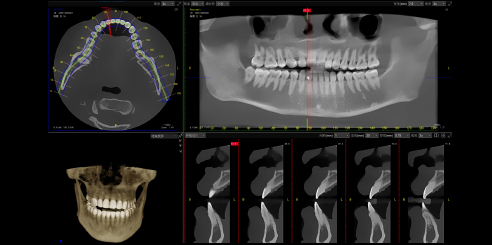
- Imaging
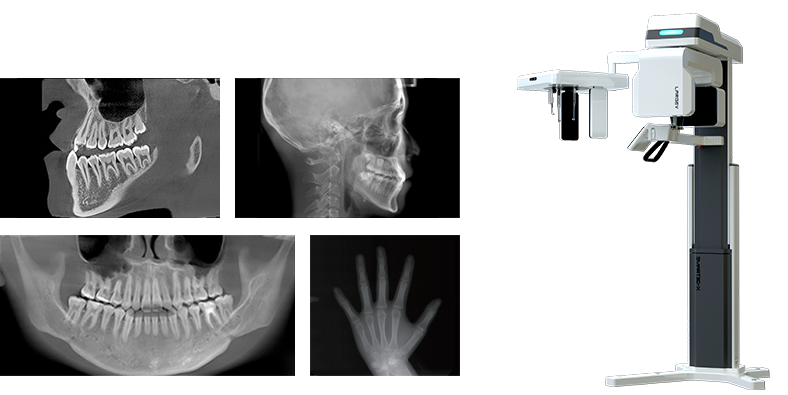
CT / CEPH / PAN
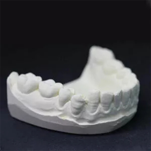
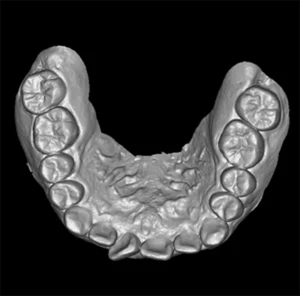
Model Scan (Optional)
Optional FOVs
Option 1
Option 2
Option 3

12x10cm

8x8cm

5x8cm


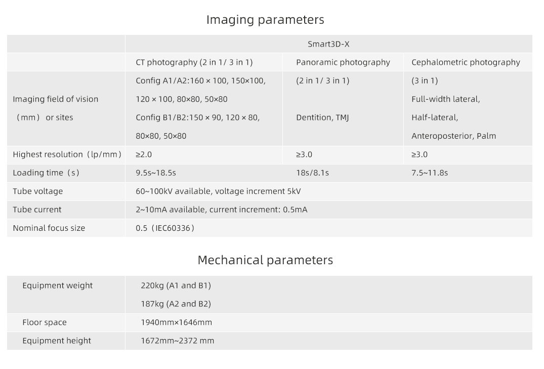
High resolution up to 2.0 lp/mm
Voxel size 0.05-0.25mm
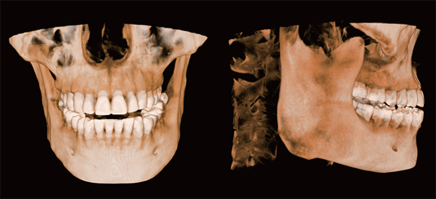
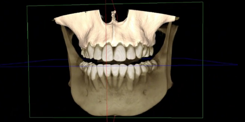
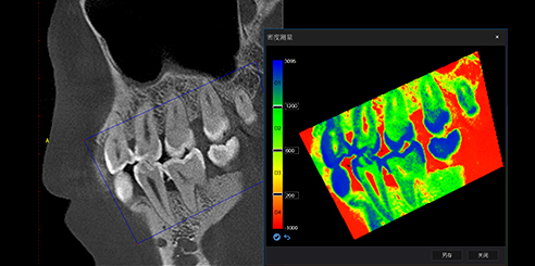
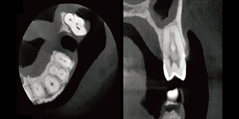
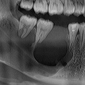
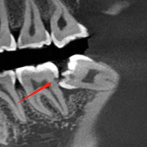
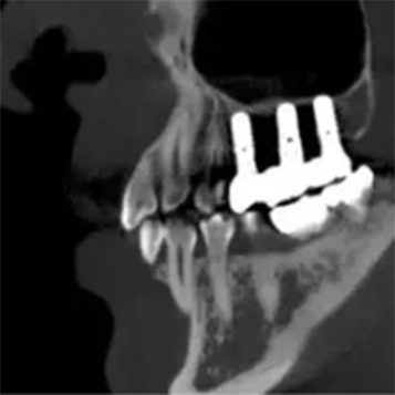
High-Definition Image - Helps Accurate Diagnosis and Treatment
0.5mm small focus tube ensures outstanding image quality.
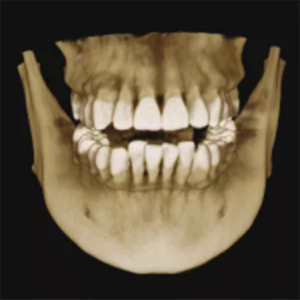
Resolution up to 2.0lp/mm, voxel size of 0.25~0.05 mm optional.
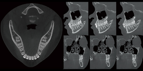
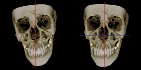
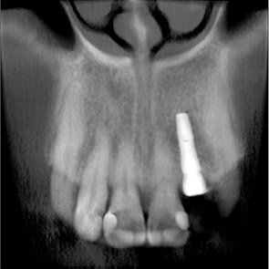
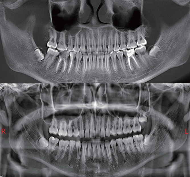
Panoramic Image Reconstructed from 3D Image Data
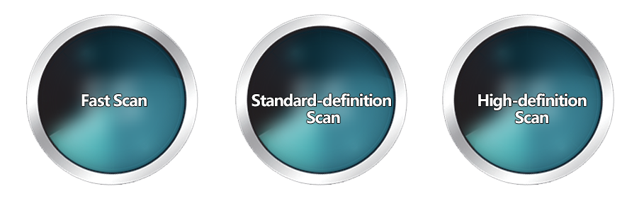
Three Scan Modes
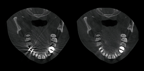
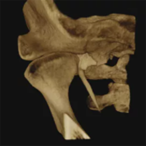
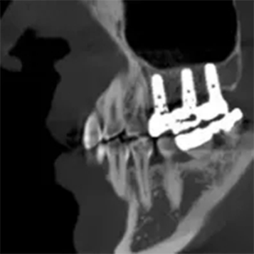
T-MAR Metal Artifact Reduction

360°scan and 800 frame images with unique CT algorithms

QuartZ 4 scan platform, supporting flexible scan mode

Multiple focus layers in panoramic imaging, fitting the patient's dental arch

Easy-to-target scan area

Six positioning lasers with face-to-face communication to posit precisely

X-type base is convenient for wheelchair-bound patients

10"LED touch screen

Storage box design

Voice reminder
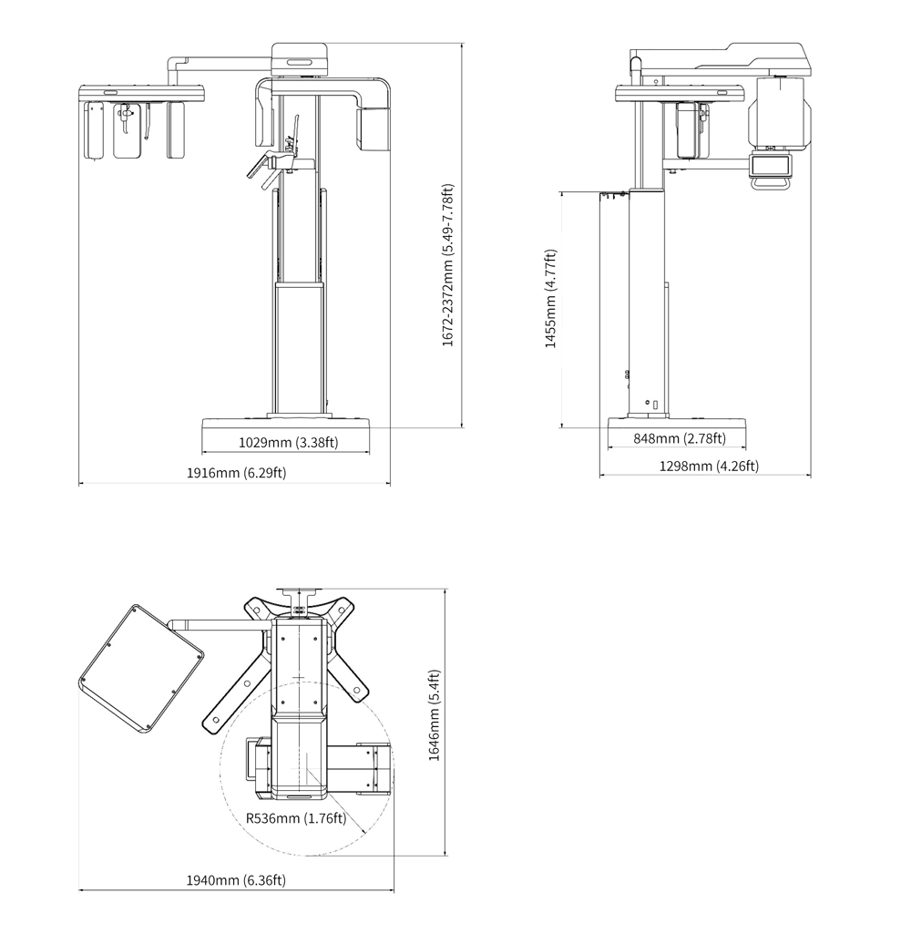
Product Size Display
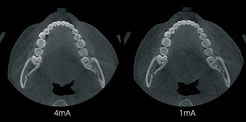
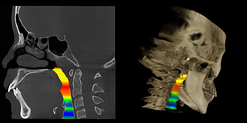
AI+Low dose
Boosted by the deep learning-based CT reconstruction algorithm, the Smart3D-X can obtain more defined tomography while further reducing the radiation dose, continuing to raise the industry standard for low-dose control.
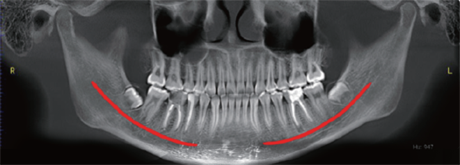
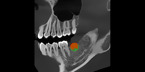
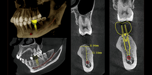
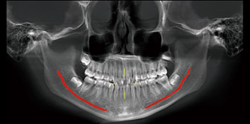
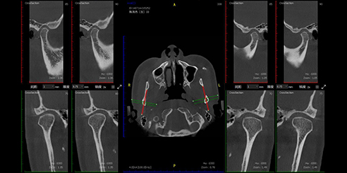
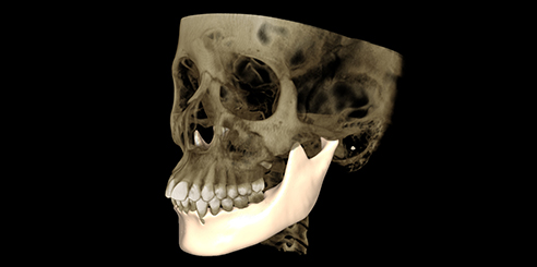
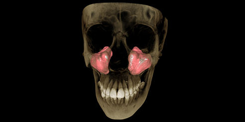
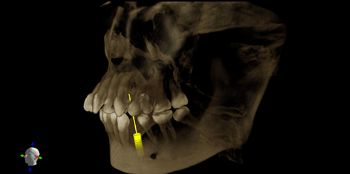
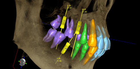
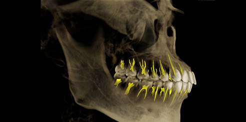
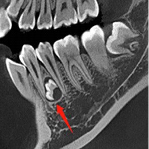
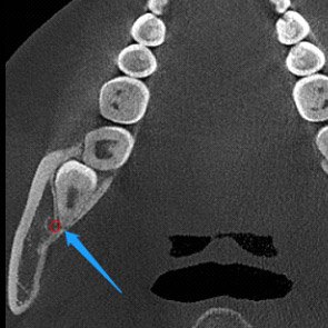
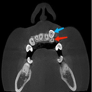
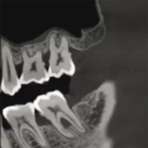
AI+Nerve
The system can label the neural tube automatically in the CT image, providing great convenience for diagnosis.
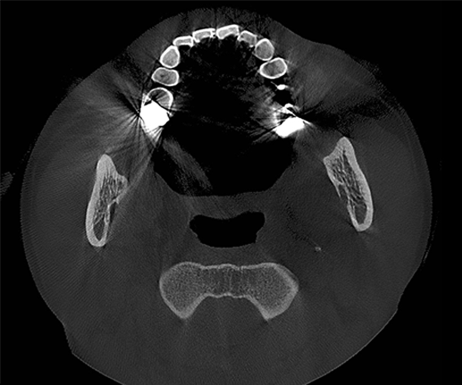
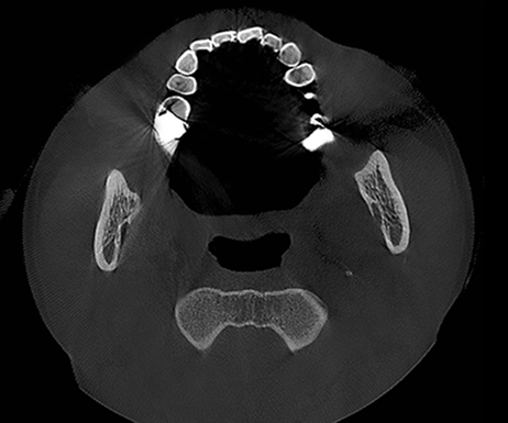
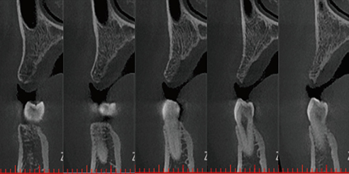
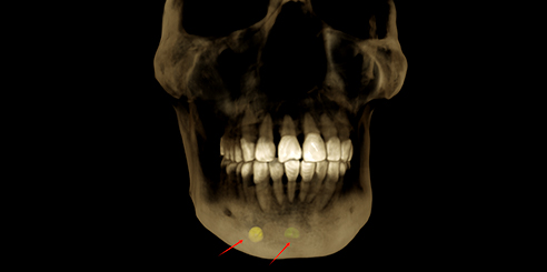
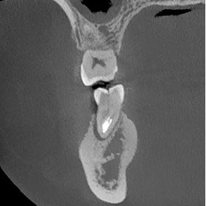
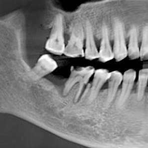
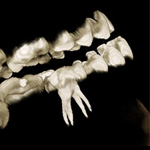
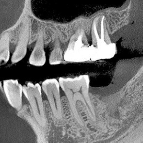
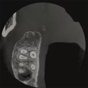
Metal Artifact Reduction
With the new T-MAR reduction module for metal artifact removal, the system corrects metal artifacts intelligently. It avoids over modification and saves the original clinical data.
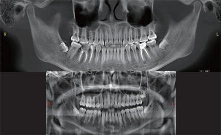
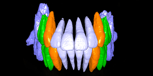
AI+PAN
CT reconstruct panoramically with the new deep learning-based CT reconstruction algorithm, the system can obtain a precise CBCT image. Panoramic images together with the new intelligent auto-focus and multilayer panoramic technology, the system automatically fits the best panoramic curves and reconstructs a better image.
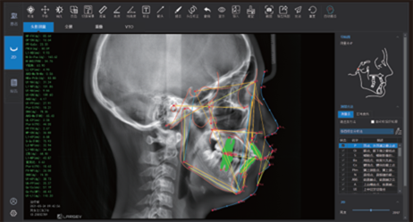
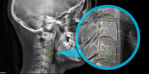
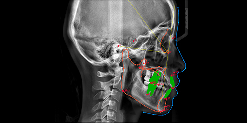
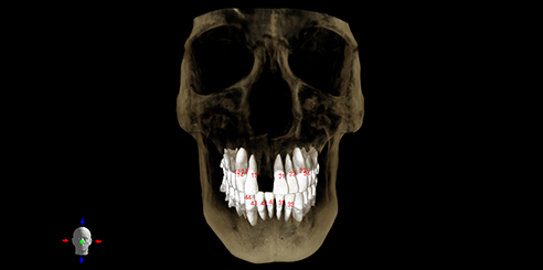
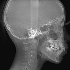
AI+CEPH Measurement (Optional)
The neural network is trained by mega data, which automatically identifies orthodontic anatomical landmark points, draws anatomical structures and outputs measurement reports according to the selected measurement methods.